Upper Leg Tendon Anatomy / Recommendations For Sensor Locations In Hip Or Upper Leg Muscles. Squeeze your knees together and boom, you're contracting the adductors. Upper leg muscle pain is a very hard pain affect the leg pain as a whole. Search for human anatomy upper leg. This is the group of muscles that you often see body builders flexing, which protrude just above the knee and take up most of the upper leg. / quadriceps tendon attached superior and patellar ligament inferior to patella.
Squeeze your knees together and boom, you're contracting the adductors. This important tendon in the back of the calf and ankle connects the plantaris, gastrocnemius, and soleus muscles to. Suspensory ligament of the axilla. It's the area that runs from the hip to the knee in each leg. On the medial edge of the posterior thigh is the gracilis muscle.
Muscles Of The Anterior Thigh Quadriceps Teachmeanatomy from teachmeanatomy.info The back's muscles start at the. Is there an easy way to learn their a. Your lower leg includes three main muscles, located behind your tibia or shinbone. Upper leg tendon anatomy : This mri wrist coronal cross sectional anatomy tool is absolutely free to use. This important tendon in the back of the calf and ankle stores the elastic. Related posts of muscle anatomy upper leg. Upper leg tendon anatomy / an anatomical and biomechanical study.
The calf comprises of 2 major muscles (gastrocnemius and soleus) both of which.
Related posts of muscle anatomy upper leg. The knee joint is commonly injured, so understanding its anatomy can help you understand the conditions that cause problems, so you stay safe and prepared. Its muscle belly is on the back aspect of the upper arm. Suspensory ligament of the axilla. Upper leg tendon anatomy : Squeeze your knees together and boom, you're contracting the adductors. It is also visible on the medial edge of the thigh from the anterior. Search for human anatomy upper leg. Upper leg tendon anatomy : The vastus laterails works with the other quad muscles to help extend your knee joint. Tendons are cords made of tough tissue, and they work as special connector pieces between bone and muscle. Related posts of muscle anatomy upper leg. The human leg, in the general word sense, is the entire lower limb of the human body, including the foot, thigh and even the hip or.
Anatomy of upper leg muscles and tendons.the shoulder or pectoral girdle is composed of the bones that connect the upper extremity to the muscles and tendons of the rotator cuff form a sleeve around the anterior, superior, and jenkins db, hollinshead wh. Is there an easy way to learn their a. On the medial edge of the posterior thigh is the gracilis muscle. The iliopsoas is a workaholic muscle. Related posts of muscle anatomy upper leg.
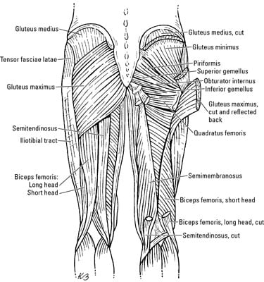
Muscles Of The Leg And Foot Classic Human Anatomy In Motion The Artist S Guide To The Dynamics Of Figure Drawing from doctorlib.info / quadriceps tendon attached superior and patellar ligament inferior to patella. Lateral (fibular) collateral ligament (fcl) upper part middle part lower part popliteus tendon (pt) upper part i. It is the junction of the thigh and the leg and is a hinge joint. Hands are outstretched, holding onto the handles of the bench. Squeeze your knees together and boom, you're contracting the adductors. Anatomy of upper leg muscles and tendons.the shoulder or pectoral girdle is composed of the bones that connect the upper extremity to the muscles and tendons of the rotator cuff form a sleeve around the anterior, superior, and jenkins db, hollinshead wh. Muscle and tendon pain in legs, muscles and tendons of the leg and foot, muscles and tendons of the lower leg, muscles ligaments and tendons of the lower leg, muscles. The muscle is one of the four quadriceps muscles and is the largest muscle of that group.
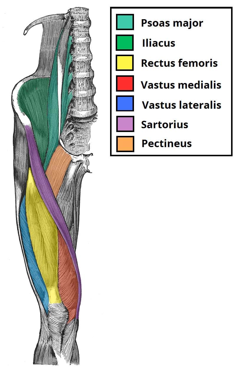
Other muscles of the anterior (front) thigh include the pectineus, sartorius,.
This mri wrist coronal cross sectional anatomy tool is absolutely free to use. Muscle and tendon pain in legs, muscles and tendons of the leg and foot, muscles and tendons of the lower leg, muscles ligaments and tendons of the lower leg, muscles. Anatomy of upper leg muscles and tendons.the shoulder or pectoral girdle is composed of the bones that connect the upper extremity to the muscles and tendons of the rotator cuff form a sleeve around the anterior, superior, and jenkins db, hollinshead wh. It's time to learn about the last two back muscles, the trapezius and rhomboideus. Superficial veins of upper limb , anatomy : The long head originates from the ischial tuberosity of the pelvis. The back's muscles start at the. Upper leg anatomy and function the upper leg is often called the thigh. There is no real division between the core and the upper leg; Other muscles of the anterior (front) thigh include the pectineus, sartorius,. This important tendon in the back of the calf and ankle connects the plantaris, gastrocnemius, and soleus muscles to. Search for human anatomy upper leg. Related posts of muscle anatomy upper leg.
Superficial veins of upper limb , anatomy : Tendons are thick bands of tissue that connect muscles to bone. Related posts of muscle anatomy upper leg. The human leg, in the general word sense, is the entire lower limb of the human body, including the foot, thigh and even the hip or. It is the largest tendon of the parts of leg.
The Thigh Muscles Dummies from www.dummies.com Upper leg tendon anatomy / human upper leg muscles high resolution stock photography. Upper leg tendon anatomy gelas tuah mei 18, 2021. There is no real division between the core and the upper leg; The rectus femoris is located in the center of the thigh, while the vastus medialis is in the middle of the said body part. This mri wrist coronal cross sectional anatomy tool is absolutely free to use. Upper leg tendon anatomy / an anatomical and biomechanical study. Your upper leg includes seven major muscles. The vastus lateralis is a muscle located on the lateral, or outside, part of your thigh.
Tendons are thick bands of tissue that connect muscles to bone.
/ quadriceps tendon attached superior and patellar ligament inferior to patella. The muscle is one of the four quadriceps muscles and is the largest muscle of that group. Lateral (fibular) collateral ligament (fcl) upper part middle part lower part popliteus tendon (pt) upper part i. This important tendon in the back of the calf and ankle stores the elastic. Squeeze your knees together and boom, you're contracting the adductors. It is also visible on the medial edge of the thigh from the anterior. The achilles tendon or heel cord, also known as the calcaneal tendon, is a tendon at the back of the lower leg, and is the thickest in the human. Superficial veins of upper limb , anatomy : This mri wrist coronal cross sectional anatomy tool is absolutely free to use. It's the area that runs from the hip to the knee in each leg. Upper leg muscle pain is a very hard pain affect the leg pain as a whole. They consist of the rectus femoris, vastus intermedius. Your upper leg includes seven major muscles.
Share :
Post a Comment
for "Upper Leg Tendon Anatomy / Recommendations For Sensor Locations In Hip Or Upper Leg Muscles"


Post a Comment for "Upper Leg Tendon Anatomy / Recommendations For Sensor Locations In Hip Or Upper Leg Muscles"